A whole-head NIRScap, measuring over 100+ channels of fNIRS data
Probes and caps are some of the most important pieces of any NIRS system setup. Both you and your subject will be in intimate contact with these critical components every time the system is used.
NIRx offers complete flexibility for montage definition. The possibilities are nearly limitless. One may place probes on the 'standard' (e.g, 10-20, 10-10, 10-5) layout or in arbitrary positions; one may use short-distance measurements (tomography) or fixed-distance measurements (topography); one may have integrated EEG or other modalities (tDCS, accelerometer, etc.) in addition to NIRS, or simply use NIRS alone on a dedicated NIRScap; and one may use fNIRS concurrently with fMRI, TMS, MEG, EKG, pulse oximetry, EMG, EOG, eye-tracking, motion-tracking, etc.
NIRS Regions of Interest
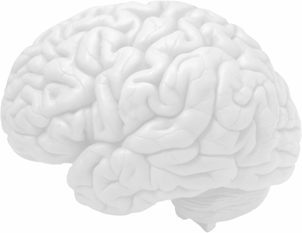
All NIRx systems allow for flexible placement of probes – data may be collected from any part of the head (actually, the probes can also be set up for peripheral fNIRS data collection as well). Here is just a sample of the areas one can easily measure with a NIRx fNIRS system:
fNIRS Brain Regions of Interest
- Pre-frontal cortex
- Dorsa-lateral pre-frontal cortex
- Supplementary motor area
- Premotor cortex
- Primary Motor cortex
- Somatosensory cortex
- Posterior parietal cortex
- Primary auditory cortex
- Broca's / Wernicke's area
- Primary visual cortex
- Others: contact NIRx
Contact us if you have questions on NIRScaps.
'Standard' vs. Arbitrary NIRS Layouts
Probes may be placed on the ‘standard’ EEG layout positions or in arbitrary positions. In either case, it is vital that one ensures proper spacing between probes, as probe distance effects light penetration and signal quality. NIRx offers hardware and software solutions that help ensure proper spacing.
NIRS layout in standard EEG 128 positions
Right: a 16-source/16-detector NIRS layout for motor measurements
Each source is red, each detector is blue, and each 'NIRS channel of interest' is purple.
In this case there are 48 NIRS channels from this 32-probe layout.
NIRS topography
The montage to the right is a 'topographic' layout, with 3cm spacing between each probe.
NIRS layout in arbitrary positions
Right: a 48-source/32-detector NIRS layout for high-density prefrontal measurements
Each source is red, each detector is green. ('NIRS channels of interest' are not shown for simplicity)
In this case there are 182 NIRS channels from this 80-probe layout.
NIRS tomography
The montage to the right is a 'tomographic' layout, with multi-distance spacing between probes to yield short and long-distance measurements.
Integrated NIRS-EEG Layouts
NIRx NIRS probes may be placed alongside EEG or other integrated cap measurement modality probes (e.g., tDCS, cochlear implants, accelerometer, etc.). It is not necessary for NIRS positions (or EEG, for that matter) to be in the standard layout. Cap montage is flexible.
The setup features a 32-channel EEG layout combined with a comprehensive whole-head fNIRS montage, consisting of a 31x31 grid and 8 short channels.
The EEG electrodes are represented by green dots, while the fNIRS includes red dots for sources and blue dots for detectors. In total, the fNIRS montage comprises 95 channels.
NIRS cap integration with other modalities
In addition to NIRS-EEG integration, NIRx offers cap integration solutions for other modalities that require access to a subject's head: NIRS-tDCS, NIRS-accelerometers, and NIRS-cochlear implants. If you would like to integrate any other sensors with a NIRScap, please get in touch.
Contact us if you have questions on NIRScaps.
Active and Passive EEG Electrode Integration
NIRx systems, probes, and caps can easily integrate with ‘active’ or ‘passive’ EEG electrodes alike.
Right: integrated NIRScap with active EEG electrodes and active NIRS optodes
Right: integrated NIRScap with passive EEG electrodes and NIRS optodes (active high-powered LEDs and fiber optic detectors)
Contact us if you have questions on NIRScaps.
NIRS optodes must be secure
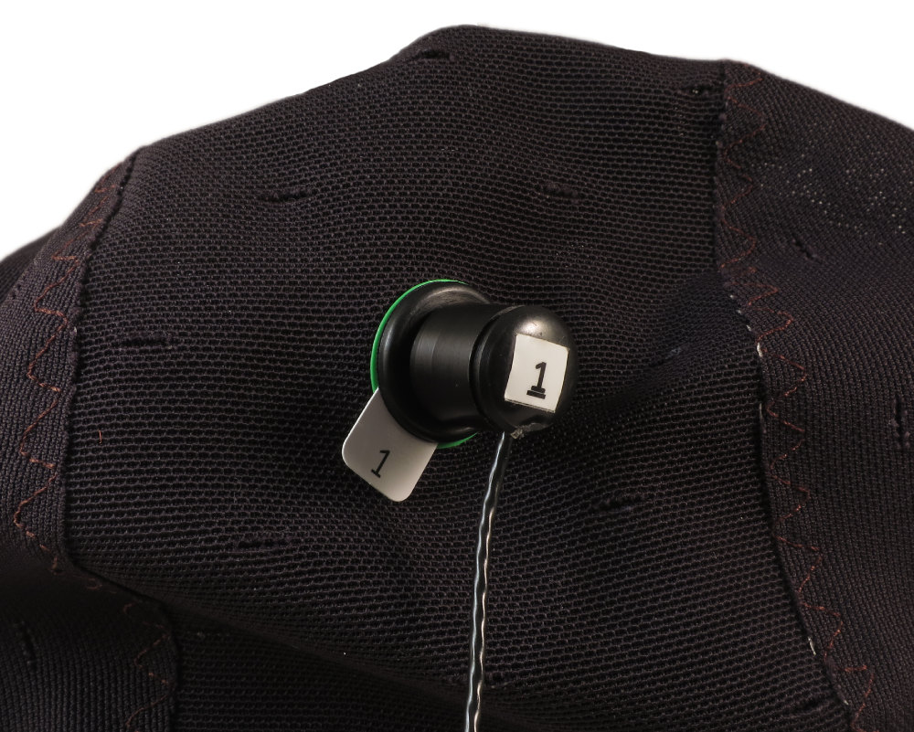
and make proper contact with the scalp
In order to send light into the brain to get a NIRS measurement of neuroactivation, one must first ensure that optodes are touching the skin.
NIRx NIRS optodes are very easy to set up incredibly quickly.
Contact us if you have questions on NIRScaps.
NIRS Probes and Caps must be fit properly and comfortably
NIRS Probes and Caps must be fit properly and comfortably Caps must be snug but comfortable. An uncomfortable subject will not hold still, or give good data. Many NIRS probes are incredibly uncomfortable and can only be worn for 30 minutes at the most before subjects must take them off. Not only are our standard probes comfortable, but we also offer specialized probes for long-term measurements and NIRS measurements on infants and children.
Standard tip NIRS optode
For all-around use on adults, juveniles, and older children.
Blunt tip NIRS optode
Ideal for use on neonatal infants to children.
Dual tip NIRS optode (premium)
More comfortable than standard tips, more sensitivity in active detectors, faster setup. (patent pending)
Dual-tipped optodes as fiber optic laser sources, fiber optic LED sources, active LED sources, fiber optic detectors, and active detectors.
The spread of the force of impact to two points makes this style of probe excellent for sensitive subjects as well as adults.
Low-profile NIRS optode (premium)
Ideal for concurrent NIRS + TMS, NIRS + MRI, or NIRS + MEG
NIRx low-profile probes lock in place, ensuring stable measurements.
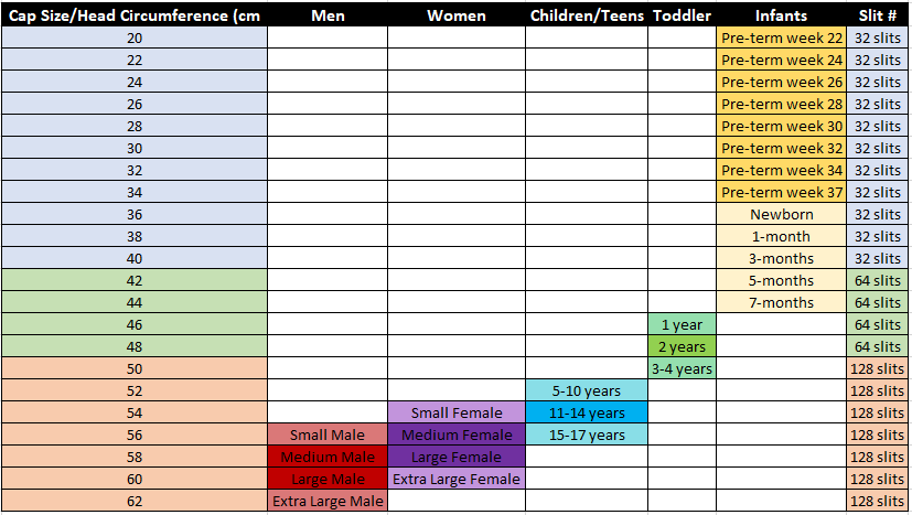
NIRx offers comfortable and properly-fitting caps for all subject ages.
NIRSCap & NIRx Probe Disinfection
For guidelines on disinfection of NIRScaps and NIRx probes, please visit this page.


